
 FP0442 : Mechanotransduction: A paradigm shift in the pathogenesis of age-related macular degeneration
Dr. SIDDHARTH NARENDRAN
Abstract
Geographic Atrophy (GA), responsible for 20% of all blindness due to AMD is characterized initially by sub-RPE deposits that accumulate between the RPE and the Bruch’s membrane (BM). The last three decades of AMD research have been primarily focused on identifying the biochemical components of these sub-RPE deposits and targeting individual components of these deposits has been the predominant treatment strategy. Despite being the pathological hallmarks of GA and AMD, the effect of the mechanical changes caused by these deposits on RPE homeostasis has not been studied. In exciting, new studies, we observed a role for mechanotransduction, a phenomenon governing the fates and functions of biological systems by mechanical forces, in RPE homeostasis and degeneration in GA. By using a gamut of mechanobiological techniques, this study provides new evidence to suggest a paradigm shift in our understanding of the pathogenesis of GA thereby identifying potential novel therapeutic strategies.
Full Text
Introduction:
GA is characterized initially by sub-RPE deposits that accumulate between the RPE and the Bruch’s membrane (BM). The last three decades of AMD research have been primarily focused on identifying the biochemical components of these sub-RPE deposits and targeting individual components of these deposits has been the predominant treatment strategy. However, these strategies thus far, have been a futile endeavor as evidenced by multiple clinical trials. Despite being the pathological hallmarks of GA and AMD, the effect of the mechanical changes caused by these deposits on RPE homeostasis has not been studied.
Mechanotransduction, a phenomenon governing the fates and functions of biological systems by mechanical forces, has been found to occur in all corners of the biological realm with an extensive and diverse repertoire of mechanisms. In exciting, new preliminary studies, we observed a role for mechanotransduction in RPE homeostasis and degeneration in GA. Hence, although inflammation is considered to be the primary process by which RPE cell death occurs in GA, we hypothesize that while RPE degeneration is perpetuated by inflammatory mediators, it is initiated by mechanical factors.
Methods:
Human tissue:
Doner eyes from patients with GA due to AMD and normal age-matched control were obtained from various eye banks. The diagnoses were confirmed by both the medical history and post mortem examination. The study followed the guidelines of the Declaration of Helsinki.
Mice
All animal experiments were performed in accordance with the Association for Research in Vision and Ophthalmology Statement for the Use of Animals in Ophthalmic and Visual Research. Mice between 6-8 wks. were used. Wild-type C57BL/6J, Best1-Cre, TAZ f/f, YAP/TAZ f/f mice were obtained from the Jackson laboratory. YAP f/f mice were bred from YAP/TAZ f/f mice. For all procedures, anesthesia was achieved by intraperitoneal injection of 100 mg/kg ketamine hydrochloride (Ft. Dodge Animal Health) and 10 mg/kg xylazine (Phoenix Scientific), and pupils were dilated with topical 1% tropicamide (Alcon Laboratories) and 2.5% phenylephrine (Alcon Laboratories).
Subretinal injection:
Subretinal injections (SRI) were performed in mice using a 35-gauge needle (Ito Co. Fuji, Japan) as described earlier. AAV1-BEST1-Cre or AAV1-BEST1-GFP were injected at 1.0×10^11 using a in Yapf/f, Tazf/f, Yap/Taz f/f mice or wild-type mice.
Fundus photography::
Photos of mice fundus were acquired by TRC-50 IX camera (Topcon) linked to a digital imaging system (Sony).
Assessment of RPE degeneration:
Sever or fourteen days after SRI, RPE health was assessed by fundus photography and immunostaining of the zonula occludens-1 (ZO-1) on RPE amount described before. Briefly, mouse RPE and choroidal flat mounts were fixed with 2% paraformaldehyde, stained with rabbit polyclonal antibodies against mouse ZO-1 and Alexa Fluor 594 conjugate (1:100, Fisher). RPE degeneration quantification was assessed by three masked graders.
Histology:
Hematoxylin and eosin staining were performed as described before []. Briefly, mice eyes were collected and embedded in Optimal Cutting Temperature Compound (Fisher) and frozen in precooled isopentane by liquid nitrogen. Cryosectioned slices at 10 um thickness were stained using the H&E Frozen section staining kit (Thermo scientific).
Cell culture:
Cell lines were cultured at 37 C and 5% CO2. Primary mouse RPE cells were isolated as previously described and grown in Dulbecco’s modified Eagle’s medium (DMEM) supplemented with 10% FBS and standard antibiotics concentrations. Primary human RPE cells were isolated as previously described and maintained in DMEM supplemented with 20% FBS and antibiotics.
Transient Transfection:
All siRNA (Silencer select) were purchased from Thermo Fisher. Human or mouse RPE cells were transfected with human Yap siRNA (s20366), mouse Yap (187076), human TAZ siRNA (Thermo Fisher), mouse TAZ siRNA (175678), and negative control (4390843) with Lipofectamine
Western blot:
Cell and tissue lysates prepared in RIPA buffer were homogenized by sonication. Protein concentration was determined with a Pierce BCA Protein Assay Kit (Thermo Fisher Scientific). Equal quantities of protein (10–50 μg) prepared in Laemmli buffer were resolved by SDS–PAGE on Novex Tris-glycine gels (Invitrogen) and transferred onto Immobilon-FL PVDF membranes (Millipore). The transferred membranes were blocked in Odyssey Blocking Buffer (PBS) or 5% nonfat dry skim milk for 1 h at RT and then incubated with primary antibody at 4 °C overnight. The antibodies used were as follows: rabbit anti-GAPDH (1:1000, cell signaling technology, 2118), mouse anti-TAZ (1:1000, Fisher Scientific, 560235), mouse anti-YAP (1:1000, Santa Cruz Biotechnology, sc-101199), rabbit anti-Yap1 (1:1000, Cell Signaling Technology, 14074).
Results:
YAP/TAZ expression is downregulated in the RPE in GA:
Western blotting (Figure 1 left, top panel), immunohistochemistry(Figure 1 left, bottom panel) and real-time polymerase chain reaction (Figure 1 right panel) show decreased YAP/TAZ expression in the RPE of donor eyes with GA.

YAP/TAZ knockdown produces spontaneous RPE degeneration:
Conditional RPE-specific knockdown of YAP and TAZ expression in the RPE induced spontaneous RPE degeneration.

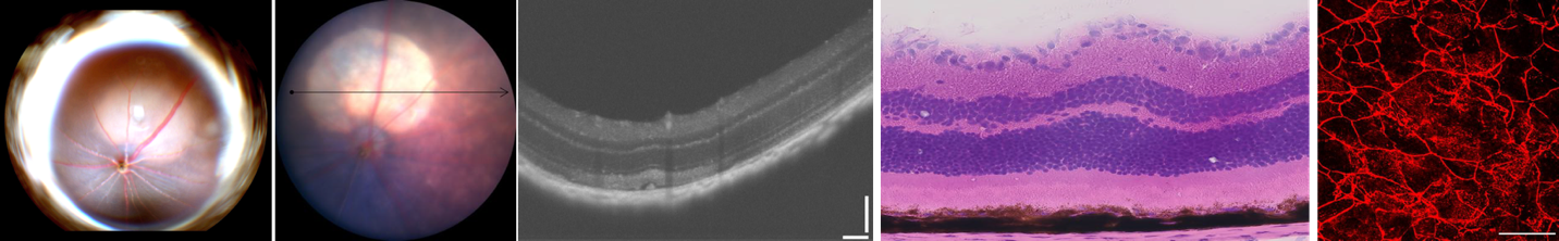
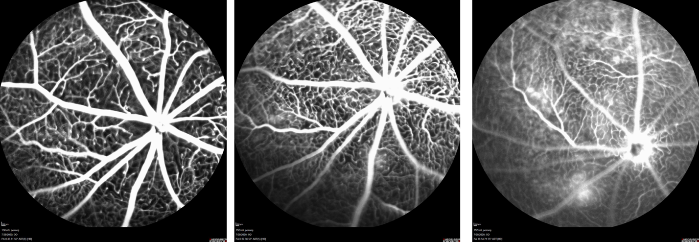
TAZ knockdown produces spontaneous choroidal neovascularization (CNV):
Conclusion
This study provides new evidence to suggest a paradigm shift in our understanding of the pathogenesis of GA by deciphering a role for the mechanical effects induced by sub-RPE deposits on the RPE thereby identifying potential novel therapeutic strategies.
Leave a Comment