
 FP0993 : Correlating the VEGF levels with the prompt clinical regression of Tubercular granulomas
Dr. MANISHA AGARWAL
Dr.Chanda Gupta
Abstract
Purpose: 10 patients with tubercular granuloma showing a prompt clinical regression with intravitreal injections of anti-VEGF bevacizumab and moxifloxacin along with ATT and oral corticosteroids.
Methods: Intravitreal injections of anti-VEGF bevacizumab and moxifloxacin given along with ATT and oral corticosteroids till complete regression of the granuloma. Aqueous sample collected was subjected to enzyme immunosorbent assay (ELISA) for analyzing intraocular VEGF-A levels by using VEGF antibody pair kit.
Results: The mean baseline VEGF level was 1004.27±411.40 pg/ml (401.32-1688.95). The mean number of intravitreal injections was 3.1 (2-4). The mean VEGF level at the time of last injection was 27.62±46.86 pg/ml (6.9-131.83).
Conclusions: Anti-VEGFs along with moxifloxacin may be beneficial as an adjunct to ATT for a prompt regression of TB granulomas. More frequent than monthly injections maybe required. due to much higher levels of VEGF in these patients.
Full Text
Aditi Dhawan, BSc, MScb, and Vivek Jha, MDa
aVitreoretina Department, Dr Shroff’s Charity Eye Hospital, New Delhi, India; bProduct Development Cell, National Institute of Immunology, New Delhi, India
Purpose: To report pre and post treatment levels of VEGF-A in the aqueous humour of patients with intraocular tubercular granulomas and study the effect of a combined intravitreal anti-VEGF bevacizumab and moxifloxacin therapy on their regression.
Methods:Aqueous samples of 10 consecutive patients with intraocular tubercular granulomas obtained before and after initiating treatment were subjected to ELISA for analysing intraocular VEGF-A levels. Intravitreal injections of bevacizumab and moxifloxacin were given weekly till com- plete regression of these granulomas. All patients received the usual four-drug ATT and oral corticosteroids.
Results: Mean baseline VEGF-A level was 1004.27±411.40 pg/ml (401.32-1688.95) that reduced significantly to 27.62±46.86 pg/ml (6.9-131.83) at the last injection. Meannumber of intravitreal injections was 3.1 (2-4). We found significant correlation of decreasing levels of aqueous VEGF-A with the clinical regression of these tubercular granulomas.
Conclusions:Anti-VEGFs along with moxifloxacin may be beneficial as an adjunct to ATT for a prompt regression of TB granulomas. More frequent than monthly injections maybe required due to much higher levels of VEGF in these patients.
Abbreviations: TB: Tuberculosis; IOTB: Intraocular tuberculosis; VEGF: Vascular endothelial growth factor; RD: Retinal detachment; Mtb: Mycobacterium tuberculosis; ATT: Antitubercular therapy; AMD: Age-related macular degeneration; SRF: Subretinal fluid; ELISA: Enzyme immunosorbent assay; PCR: Polymerase chain reaction; ONH: Optic nerve head; MDR-TB: Multidrug-resistant tuberculosis; pg/ml: picogram/milliliter; ESR: Erythrocyte sedimentation rate; CECT: Contrast enhanced computed tomography; DNA: Deoxyribonucleic acid; RNA: Ribonucleic acid; BSL: Biosafety level; BCVA: Best corrected visual acuity; HM: Hand movements; KP: Keratic precipitates; PSC: Posterior subcapsular cataract; PS: Posterior syne- chiae; CRA: Chorio-retinal atrophy; IVMP: Intravenous methyl prednisolone; OCT: Optical coherence tomography; RPE: Retinal pigment epithelium; FFA: Fundus fluorescein angiography; ICG: Indocyanine angiography; RAP: Retinal arterial proliferans.
Tubercular granulomas are the most well recognized pheno- type of intraocular tuberculosis (IOTB), and present as yellow- ish subretinal elevated lesions often accompanied by an exudative retinal detachment (RD).1 In experimental models of IOTB, these granulomas were found to show overexpression of vascular endothelial growth factor (VEGF).
It has been proposed that increased levels of VEGF may be responsible for the vascularization of these granulomas.3,4 Moreover, pharmacological inhibition of the VEGF pathway may limit the spread of Mycobacterium tuberculosis (Mtb) infection causing regression of the new vessels, and thus forms the basis of newly emerging concepts in considering host directed therapies over the usual pharmacological anti- tubercular therapy (ATT) that has been the standard of care for over six decades.
The use of ATT and oral corticosteroids in the management of tubercular granulomas has been well documented in the past.6 In recent years, a number of case reports have reported successful use of anti-VEGF therapy along with the standard care in the treatment of tubercular granulomas.7–12
To the best of our knowledge, pre- and post-treatment levels of VEGF-A in the aqueous humour of patients with tubercular granulomas have not been studied. We report pre and post treatment results of VEGF-A levels in 10 consecutive patients with intraocular tubercular granulomas who showed a prompt regression with weekly intravitreal injections of anti-VEGF bevacizumab and moxifloxacin along with the usual standard of care ATT and oral corticosteroids. A correlation was done of their clinical regression with the intraocular aqueous levels of VEGF-A.
Material and methods
All consecutive patients between January 2017 and January 2020 who presented with intraocular tubercular granulomas at the uvea clinic of a tertiary eye care hospital in North India were included in the study. All these patients met the diagnos- tic criteria for IOTB according to the classification by Gupta et al.13 The institutional ethics committee approval was obtained and the study adhered to the tenets of the Declaration of Helsinki. An informed written consent was obtained from all the patients. A detailed history of any exposure to pulmonary tuberculosis (TB) was taken. All patients underwent complete hemogram with erythrocyte sedimentation rate (ESR), Mantoux test, contrast enhanced computed tomography (CECT) of the chest and other relevant laboratory investiga- tions to rule out other infectious etiologies. A thorough sys- temic examination was carried out by an in-house physician.
Optical coherence tomography (OCT) and color fundus photo were done at baseline and after every injection. Fundus fluor- escein angiography (FFA) and indocyanine green angiography (ICGA) were performed at the discretion of the treating clin- ician. All the patients were treated with intravitreal injection of anti-VEGF bevacizumab (1.25 mg/0.05 ml; off label use) and moxifloxacin (500 μg/0.1 ml; off label use) under topical anesthesia and aseptic precautions, along with four drug ATT and oral corticosteroids (1–1.5 mg/kg/day). Weekly intravitreal injections were continued till there was a clinical regression of the granulomas and complete resorption of the subretinal fluid (SRF) on OCT. Complete regression of the granuloma was defined as flattening of the granuloma with normalization of the contour of the overlying retinal layers with or without chorio-retinal atrophy (CRA) and resorption of the overlying SRF. Patients who were already receiving ATT therapy for pulmonary or extrapulmonary TB were not excluded from the study.
Anterior chamber paracentesis of the aqueous fluid (0.1 ml) was done immediately after injecting the drugs, thereby nor- malizing the intraocular pressure. The aqueous fluid sample thus collected in a sterile syringe was packed, labelled and then transported in an insulated box with dry ice to the laboratory. The samples were subjected to sandwich enzyme immunosor- bent assay (ELISA) for analyzing intraocular VEGF-A levels. VEGF-A levels were calculated by using human VEGF anti- body pair kit {Invitrogen; 10 plate Format; Lot#*: 650073; Catalog#CHG0113}. The linear range of detection was 2– 2000 pg/mL (picograms/milliliter).Polymerase chain reaction (PCR) for the Mtb genome was performed in five patients (anterior chamber sample in four and vitreous sample in one). PCR was not done in the other five patients as four were already receiving ATT at the time of presentation and one patient had financial constraints. A positive Mantoux test, PCR detecting Mtb genome and findings on CECT chest helped in ruling out sarcoidosis.
Extraction of DNA from Patient Sample
Vitreous and aqueous samples were analyzed for Mtb genome detection. Initial sample handling was conducted in BSL-3 facility and the entire DNA was isolated using MasterPure™ Complete DNA and RNA Purification Kit (Epicentre-Lucigen, Cat No. MC85200). DNA was also isolated from M. tubercu- losis, H37Rv strain, culture. The yield and quality (ratio of absorption at 260 and 280 nm) of DNA product was measured using NanoDrop™ One (Thermo Fisher Scientific, ND- ONE-W4).
Detection of M. Tuberculosis DNA Using Real-Time PCR and TaqMan Probes
Two probes were designed (Table 1) for detection of M. tuber- culosis gene markers, i.e. MPB 64 (NC_018143; Rv1980c) and IS6110 (X52471). The probes were conjugated with 5ʹ-FAM and 3ʹ-TAMRA. A real-time PCR reaction (20 μL, microliter) was setup using Takyon™ Rox Probe MasterMix dTTP Blue (Eurogenetec, Cat. No. UF-RPMT-B0701), 250 nanometre (nm) probe, 300 nm forward and reverse primers, and 5 μL DNA. The reaction was performed in Mastercycler® ep realplex4 (Eppindorf AG Hamburg, Cat. No. 6302 010760). The following thermal cycling specifications were performed 3 minutes at 95°C and 40 cycles each for 10 seconds at 95°C, 45 seconds at 53°C (Mpb64)/61°C (IS6110), and 30 seconds at 72°C. All reactions were run in duplicate or triplicate form. The DNA was diluted so that 5 μL contained 102–106 copies. A standard curve was plotted between the copy number and control value from real-time PCR to calculate the bacterial load in patient samples.
Results
A total of 10 eyes of 10 patients were included in the study. Eight patients were females and 2 patients were males. The mean age of presentation was 25.9 ± 12.27 years (5–51 years). There was a past history of pulmonary tuberculosis in 6 out of 10 patients. Left eye was affected in 7 out of 10 patients. Anterior uveitis (SUN classification)14 was present in 6 patients. Varying grades of vitritis was found in all the patients. Eight patients presented with a choroidal granuloma and two patients with optic nerve head (ONH) granuloma. Multiple patches of choroiditis extending till the equator were noted along with ONH granuloma in one patient. Exudative RD with SRF was found in 8 patients. Eight patients reported a positive Mantoux test. CECT chest was suggestive of active pulmonary TB in seven patients. PCR detected Mtb genome in four aqu- eous samples and one vitreous sample. Four drug ATT was started in five patients and in the other five patients the ongoing ATT was continued, for a minimum duration of 9 months (9–12 months) (Table 2).
Table 1. Primer and probe design.
| S. no. | Gene markers | Probe | Primers |
|---|---|---|---|
| 1 | MPB64 | 5ʹ-CCTACAACATCAACATCAGCCTGC-3’ | Forward: 5ʹ-GTGCCAGATTCAAATGTC-3’ Reverse: 5ʹ-GGTGATATTCAATTCGTAGG-3’ |
| 2 | IS6110 | 5ʹ-TGTGCTCCTTGAGTTCGCCA-3’ | Forward: 5ʹ-CCTACTACGACCACATCA-3’ Reverse: 5ʹ-CCGTAAACACCGTAGTTG-3’ |
Table 2. Clinical profile of patients with tubercular granuloma.
| Case no. | Age (yrs) | Sex | Eye | H/o TB | Anterior segment | Posterior segment | Baseline BCVA | Final BCVA | Mantoux (mm) | CECT Chest | PCR | ATT | Corticosteroid | No of injections |
|---|---|---|---|---|---|---|---|---|---|---|---|---|---|---|
| 1 | 22 | F | Left | Yes | Quiet | Vitritis, Choroidal granuloma, exudative RD | 6/36, N12 | 6/6, N6 | 16×18 | Active Koch’s | Not done | Yes | Oral | 2 |
| 2 | 33 | M | Left | No | Quiet | Vitritis, Choroidal granuloma | 6/36, N24 | 6/6, N6 | 1×1 | Normal study | Not done | Yes | Oral | 2 |
| 3 | 22 | F | Right | No | Quiet | Vitritis, Choroial granuloma | 6/9, N6 | 6/6, N6 | 2×2 | Normal study | Positive, Aqueous sample | Yes | Oral | 2 |
| 4 | 5 | F | Left | No | Flare+2, cells+2 | Vitritis, ONH granuloma, exudative RD, | HM | 6/24, N12 | 18×20 | Active Koch’s | Positive, Aqueous sample | Yes | Topical, Oral | 4 |
| 5 | 51 | F | Left | Yes | Flare +1, cells +1, KP’s, | Vitritis, ONH granuloma, exudative RD | 3/60, N36 | 6/18, N18 | 20×25 | Active Koch’s | Positive, Aqueous sample | Yes | Topical, Oral | 3 |
| 6 | 28 | F | Right | No | Flare +1, cells+1, 360 | Vitritis, Choroidal granuloma, exudative RD | HM | HM | 32×35 | Normal study | Positive, Vitreous | Yes | Topical, Oral | 4 |
| 7 | 17 | F | Left | Yes | Flare+1, cells+1 | Vitritis, Choroidal granuloma, exudative RD | HM | HM | 22×22 | Active Koch’s | Not done | Yes | Topical, Oral | 4 |
| 8 | 22 | F | Left | Yes | Quiet | Vitritis, Choroidal granuloma, exudative RD | 6/60, N36 | 6/18, N12 | 20×20 | Active Koch’s | Not done | Yes | Oral | 3 |
| 9 | 23 | F | Left | Yes | Flare+2, cells+2 | Vitritis, Choroidal granuloma, exudative RD | HM | 6/6, N6 | 20×22 | Active Koch’s | Not done | Yes | Topical, Oral | 3 |
| 10 | 36 | M | Right | Yes | Flare+2, cells+2 | Vitritis, Choroidal granuloma, exudative RD | HM | 3/60, N36 | 28×30 | Active Koch’s | Positive, Aqueous sample sample | Yes | Topical, Oral | 4 |
TB-Tuberculosis, BCVA-Best corrected visual acuity, CECT-Contrast enhanced computed tomography, PCR-Polymerase chain reaction, ATT-Anti-tubercular therapy, ONH- Optic nerve head, RD-Retinal detachment, HM-Hand movements, KP-Keratic precipitates, PSC-Posterior subcapsular cataract, PS-Posterior synechiae, F-Female, M-Male
At baseline presentation, 9 patients had best corrected visual acuity (BCVA) <6/36 (snellen’s chart) and one patient with 6/9. At the last follow-up, four patients had BCVA 6/6, two with 6/ 18 and four patients < 6/24. The mean duration of follow-up was 13.8 ± 7.0 months (9–24). At the last follow-up, complete regression of the granuloma with CRA and resorption of the SRF was seen in all the patients.
Case 1 had a paradoxical worsening after starting ATT and was treated with intravenous methyl prednisolone (IVMP) following which complete regression of the granuloma was seen. Case 7 developed total rhegmatogenous RD for which pars plana vitrectomy with silicone oil injection was performed.
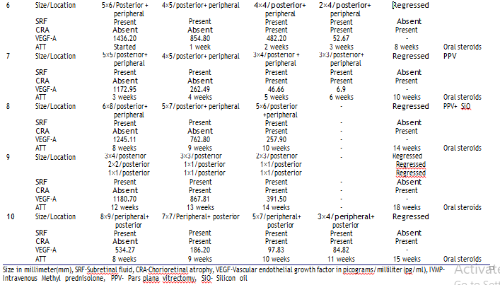
The mean baseline VEGF-A level was 1004.27 ± 411.40 pg/ml (401.32–1688.95). The mean VEGF-A level at the time of last injection was 27.62 ± 46.86 pg/ml (6.9–131.83). A significant decrease in the VEGF-A level was found with successive intravi- treal injections. Table 3 shows the correlation of the VEGF-A levels with the clinical response of tubercular granulomas.
Case reports
Case 1
A 24-year-old female presented with diminution of vision in the left eye for last 5 days. She was diagnosed with pulmonary TB and was on ATT for the last 2 months. BCVA in the right eye was 6/6, N6 and in the left eye 6/ 36, N12. Right eye fundus was within normal limits. Left eye fundus showed a large choroidal granuloma measuring3 × 6 mm in the superior quadrant with intraretinal hemorrhage and SRF reaching till the macula. The disc was hyperemic with surrounding granuloma and a small 1 × 1 mm choroidal granuloma was noted inferotemporal to the disc (Figure 1). OCT through the macula showed SRF and scan through the superior granuloma showed a choroidal bump with SRF and intraretinal cystic spaces.
Mantoux test was positive (16x18mm). CECT chest showed fibro-parenchymal lesion in the left upper lobe and calcified lymph nodes bilaterally suggestive of active Koch’s. ATT was continued and oral corticosteroids (1.5 mg/kg/day) were added. An intravitreal injection of bevacizumab and moxifloxacin was given. Baseline VEGF-A was analyzed as927.56 pg/ml. At 2-weeks follow up, extensive CRA was seen in the area of previously noted granuloma. However, a new granuloma was seen at the temporal edge of the CRA corresponding to the healed superior granuloma, extending into the macular area and threatening the fovea.
This was diagnosed as a paradoxical reaction and treated with IVMP 1 gm/kg for 3 days followed by high dose oral corticoster- oids (2 mg/kg/day) along with a repeat intravitreal injec- tion. VEGF-A level was analyzed as 422.16 pg/ml after thefirst intravitreal injection. Subsequent follow-up at 2 weeks, BCVA in the left eye improved to 6/6, N6. Fundus showed mild disc hyperemia, complete regression of the granuloma along with CRA and pigmentation and few hard exudates at the macula. OCT showed a normal foveal contour with complete resorption of the fluid.
Case 2,3
Case 2 (33 years, male) and case 3 (22 years, female) presented with uniocular diminution of vision. Case 2 had an exposure of pulmonary TB. In both the patients, on fundus examination there was presence of a choroidal granuloma with vitritis along with perivasculitis in case 3. Mantoux test was negative and CECT chest was normal in both the patients. PCR of the aqueous sample was positive for Mtb genome for case 3 and was not done for case 2 due to financial constraints. Both the cases were diagnosed as possible ocular TB.
ATT and oral corticosteroids (1 mg/kg/day) were started for both the patients by our in-house physician. Two intravitreal injections of bev- acizumab and moxifloxacin were administered at a weekly interval. Baseline VEGF-A level in case 2 and 3 was 1688.95 pg/ml and 401.32 pg/ml which decreased to 1140.70 pg/ml and 176.74 pg/ml respectively after one intravi- treal injection. In case 2, the choroidal granuloma completely melted away and patient gained BCVA of 6/6, N6 (Figure 2). In case 3, the patient gained BCVA of 6/6, N6 and regression of the granuloma was seen with CRA and pigmented scarring and hard exudates at the macula (Figure 3).
Case 4
A 5-year-old girl presented with diminution of vision in the left eye for the last 2 months. She gave a history of cough with expectoration. BCVA in the right eye was 6/6, N6 and left eye was hand movements (HM) close to face. Fundus examination of the left eye showed a large granuloma infiltrating the optic nerve head with multiple patches of choroiditis extending till the equator (Figure 4). Mantoux test was positive (18×20 mm) and CECT of the chest showed bilateral multiple ill-defined centri- lobular nodules with surrounding ground glass opacification and multiple necrotic discrete to conglomerate mediastinal and hilar lymph nodes suggestive of active pulmonary Koch’s.
She was started on four drug ATT along with oral corticosteroids (1 mg/ kg/day). PCR was positive for Mtb genome. Four intravitreal injections of bevacizumab and moxifloxacin were administered on a weekly interval. Baseline VEGF-A level was 667.82 pg/ml. It decreased to 376.78 pg/ml, 224.30 pg/ml, and 131.83 pg/ml, respectively, after successive intravitreal injections. The ONH granuloma was found to regress with each injection. Follow-up at 9 months on the completion of ATT, her vision improved to 6/24, N12. Fundus showed mild pallor of the disc with peripa- pillary CRA and complete regression of the granuloma.
Case 5
A 51-year-old female complained of diminution of vision in both the eyes for the last 3 months. She had a history of pulmonary TB fifteen years back for which she had taken ATT. BCVA in the right eye was 6/9, N6 and in the left eye 3/ 60, N36. Anterior segment of the right eye showed old keratic precipitates (KP). Left eye showed cells+1, flare+1, mutton fat KP’s, and posterior subcapsular cataract. Fundus of the right eye showed peripapillary CRA with pigmented scarring and the left eye showed a large granuloma infiltrating the ONH with intraretinal hemorrhage and peripapillary serous retinal detachment involving the macula (Figure 5). OCT through the disc confirmed a granuloma with surrounding SRF detach- ing the macula.

Mantoux test was positive (20×25 mm) and CECT chest showed a focal area of subsegmental collapse with pulled up major fissure in the apical-posterior segment of the left upper lobe suggestive of old Koch’s and a small focal area of fibro-parenchymal lesion in the apical segment of the right upper lobe suggestive of active Koch’s. PCR was positive for Mtb genome. She was started on four-drug ATT, oral corticos- teroids (1.5 mg/kg/day) and topical prednisolone acetate eye drops (1%) with atropine (1%). Three intravitreal injections of bevacizumab and moxifloxacin were administered at a weekly interval. Baseline VEGF-A level was 787.91 pg/ml which decreased to 272.02 pg/ml and 156.87 pg/ml subsequently after repeat injections. Complete regression of the granuloma with resorption of the SRF was noted. OCT showed a normal contour of the disc and the macula.
Figure 1. Case 1: (a) Left eye fundus photo showing a large choroidal granuloma 3 × 6 mm (red arrow), hyperemic disc with surrounding granuloma (green arrow) and a 1 × 1 mm choroidal granuloma inferotemporal to the disc (yellow arrow). (b) OCT macular scan showing intraretinal cystic spaces (yellow arrow) and subretinal fluid (red arrow). (c) OCT scan through the superior granuloma showing choroidal bump with intraretinal cystic spaces (yellow arrow) and subretinal fluid (red arrow). (d) Fundus photo showing regression of the granuloma (yellow arrow) 2 weeks after the first injection with a new lesion due to paradoxical worsening (red arrow). (e) OCT macular scan showing subretinal fluid (yellow arrow). (f) OCT scan through the new lesion showing choroidal bump (yellow arrow) with subretinal fluid (red arrow). (g) Fundus photo showing completely regressed granuloma (red arrow) with hard exudates at the macula (green arrow) after IVMP and second injection. (h) OCT macular scan showing normal foveal contour (i) OCT scan through the granuloma showing normal choroidal structure.

Figure 2. Case 2: (a) Left eye fundus photo showing a 3 × 6 mm choroidal granuloma in the superior quadrant (red arrow). (b and c) Fundus photo showing melting of the granuloma after the first and second injections, respectively. (d) Fundus photo showing complete disappearance of the granuloma at 9-month follow-up.

Figure 3. Case 3: (a) Right eye fundus photo showing a 3 × 4 mm choroidal granuloma along the inferior arcade (red arrow). (b and c) Fundus photo showing regressing granuloma (red arrow) along with hard exudates at the macula (yellow arrow) after the first and second injections, respectively. (d) Fundus photo showing regressed choroidal granuloma (red arrow) at 9-month follow-up.

Figure 4. Case 4: (a) Left eye fundus photo showing ONH granuloma (red arrow) with surrounding choroiditis (green arrow) and peri-vasculitis (yellow arrow). (b–e) Fundus photo showing regression of the granuloma with resolving choroiditis and peri-vasculitis after the first, second, third, and fourth injections, respectively. (f) Fundus photo showing completely regressed ONH granuloma with resolving perivasculitis (yellow arrow) at 3-month follow-up. (g) Fundus photo at 9-month follow-up showing normal ONH contour (red arrow).

Figure 5. Case 5: (a) Left eye fundus photo showing an ONH granuloma with intraretinal hemorrhage (yellow arrow). (b) OCT scan through the ONH showing granuloma (yellow arrow) with surrounding subretinal fluid (red arrow). (c) OCT macular scan showing subretinal fluid (yellow arrow). (d) Left eye fundus photo showing complete regression of the granuloma after three injections. (e) OCT scan showing normal ONH contour. (f) OCT macular scan showing complete resorption of the subretinal fluid.

Discussion
In our series of consecutive 10 eyes of 10 patients with intrao- cular tubercular granulomas, in addition to the usual ATT and oral corticosteroids, weekly injections of anti-VEGF bevacizu- mab and moxifloxacin led to a prompt regression of these granulomas that included eight choroidal and two ONH gran- ulomas. Five of these patients, who had not received prior ATT, in whom aqueous humour was subjected to PCR showed the presence of the Mtb genome in the intraocular fluid, con- firming the tubercular etiology of these granulomas.13 We also found very high levels of VEGF-A in the pretreatment aqueous humour. The VEGF-A levels decreased substantially in these eyes following the weekly intravitreal therapy and correlated significantly with the regression of the granulomas. We thus provide evidence for the rational use of the anti-VEGF therapy in the management of intraocular tubercular granulomas.
The monthly use of anti-VEGF therapy currently the gold standard for neovascular age-related macular degeneration (AMD) and macular edema due to diabetic retinopathy and retinal vascular occlusion is based on the presence of VEGF in the aqueous/ vitreous humour of these patients.15 However, we observed much higher VEGF-A levels in tubercular granulomas than have been reported in the aforementioned retinal diseases. Hence, we feel that a much higher frequency of the anti- VEGF therapy was required in our patients. The progressive decrease in the VEGF levels following weekly injections has thus justified this approach.
VEGF is a known biomarker for active TB disease in pul- monary as well as extrapulmonary sites, to monitor disease severity, bacterial burden, and therapeutic responses in pul- monary TB.16,17 Matsuyama et al16 analyzed the serum VEGF levels (722.6 ± 362.2 pg/ml) in patients with active pulmonary TB and found them significantly higher than in old treated TB patients (185.1+158.4 pg/ml). The highest serum VEGF level was 1300 pg/ml which decreased to <100 pg/ml, 6 months after ATT. They showed that the serum VEGF levels of patients with active pulmonary TB decreased parallel to improvement of the disease.
A vision threatening paradoxical worsening of IOTB due to an immunological reaction is a major fear of starting ATT which requires stepping up of oral corticosteroids and at times needs intravenous corticosteroid therapy.18 This is well demonstrated by case 1 of our series. However, Jain et al.9 have reported a case of paradoxical worsening in a HIV-infected patient with tubercular granuloma that did not respond to oral corticosteroids but showed an excellent response to two injec- tions of bevacizumab. Agarwal et al.7 also reported a case in whom even after 3 months of ATT there was no regression of the granulomas which was associated with an exudative RD. However, three weekly injections of bevacizumab and moxi- floxacin led to a complete regression of the granulomas with resolution of the exudative RD.
In the last decade, a few case reports have been published highlighting the use of intravitreal anti-VEGF agents as mono- therapy or as adjunct to ATT and oral corticosteroids for treating tubercular granulomas.7–12 Bansal et al.11 and Babu et al.12 each treated a case of vascularized tubercular granuloma with monthly intravitreal injections of bevacizumab along with ATT and corticosteroids leading to regression of the granu- loma and resolution of exudative RD. Bansal et al.11 in their case have also shown a prompt regression of the recurrent granuloma which occurred after one year of stopping ATT and was treated with two bevacizumab injections alone thereby maintaining the remission of the granuloma. Invernizzi et al.10 have reported the first case of an ONH tubercular granuloma with surrounding SRF successfully treated with monthly injec- tions of bevacizumab along with ATT and oral corticosteroids.
Jain et al.8 have reported complete regression of a vascularized tubercular granuloma with a single intravitreal injection ofranibizumab without adding ATT as they did not find any evidence of systemic TB. In contrast, we started ATT without any evidence of systemic TB in cases 2 and 3 based on the clinical signs, as suggested by Gupta et al.13 Ocular TB is a form of extrapulmonary TB presenting as uveitis in TB-endemic countries like India.1 The classic his- topathological finding in IOTB is epithelioid cell granuloma with a central area of caseation necrosis.
1 Datta et al.19 have shown that in both human and rabbit tubercular granulomas, VEGF is most highly expressed in the inner macrophage layer that surrounds the necrotic core. They also noted that vascu- lature, with the highest density in the periphery of these granulomas, was both structurally, morphologically, and functionally abnormal. Further they have shown that bevaci- zumab normalizes the granuloma vasculature, reduces hypoxia, and also enhances the small molecule delivery of anti-TB drugs.
A multidrug therapy regimen of ATT should not logically allow multidrug resistant TB (MDR-TB) to emerge. Yet, it is a well phenomenon in pulmonary TB and other extrapulmonary sites. Moreover, reports of MDR-TB are emerging in ophthal- mic literature and posing a major therapeutic challenge that calls for innovative host directed therapies.20–24 Of all the drugs that are used to treat TB, isoniazid is the only bactericidal drug which blocks the cell wall synthesis of the dividing Mtb, and its efficacy is determined directly by the rate of the multiplication of Mtb.25 Also, isoniazid is not effective against non-multi- plying or dormant Mtb. Wayna et al.26 demonstrated that Mtb stops multiplying and goes into dormancy in hypoxic conditions.
It has been proposed that one of the main factors in the emergence of MDR-TB may be the abnormal vessels that do not allow the ATT drugs to diffuse equally in all the parts of the granuloma. This effectively reduces the ATT to a monotherapy thereby enhancing the chances of developing MDR-TB. Using anti-VEGF drugs such as bevacizumab ensures equal distribu- tion of all the therapeutic agents in the granuloma significantly reducing the chances of MDR-TB.19The anti-TB drugs were introduced for the treatment of TB without knowing the pharmacokinetics and their differ- ential concentration in various cellular components of the tubercular granuloma. The rationale for using multiple drugs in ATT is due to the action of various drugs on different phases of Mtb.25 Tubercular granulomas show a favourable but slow response when treated with ATT.27 There have been reports of rapid diminution of vision with early onset of intraretinal and subretinal neovascular- ization due to arteriovenous communication leading to the formation of retinal arterial proliferans (RAP) in these granulomas.28,29 This may lead to a painful blind eye requiring a removal while being on ATT.30
In recent years, use of high-performance liquid chroma- tography coupled to tandem mass spectrometry in rabbit model of TB has shown that compared to the plasma levels, moxifloxacin is preferentially accumulated in the macro- phages with a high intracellular to extracellular ratio com- pared to other anti-TB drugs like isoniazid, rifampicin, and pyrazinamide.25 However, it diffuses poorly into the necro- tic center of the granuloma which can be penetrated only by pyrazinamide. Therefore, we used intravitreal moxiflox- acin to target the most active component of the tubercular granuloma, that is activated macrophages harboring Mtb to facilitate a quick elimination of the organisms.
Our case series differs in the following ways from what has been previously reported, firstly we gave a combination of intravitreal anti-VEGF bevacizumab and moxifloxacin injec- tion in comparison to only anti-VEGF injections used in the past. Secondly, previous case reports gave monthly anti-VEGF injections in the management of tubercular granulomas, whereas we gave weekly injections in all our patients. The rationale for repeating the anti-VEGF injection every week till the regression of the granulomas was the five times higher mean baseline intraocular VEGF-A level-1004.27 ± 411.40 pg/ ml (401.32–1688.95) as compared to the mean baseline VEGF- A level-179.7 ± 88.4 pg/ml (74.5–521.6) in patients with neo- vascular AMD,31 and that the half-life of intravitreal bevacizu- mab ranges between 2.5 and 7.3 days in non-vitrectomized eyes that may be further shortened due to the breakdown of the blood retinal barrier secondary to the inflammation leading to early washout of the drugs.32 Thirdly, we have also correlated the clinical regression in each case with the corresponding decreasing levels of the VEGF-A, which has never been reported earlier.
It is all but intuitive that efforts should be made not only to restore the vision of the patient quickly but also normalize the vasculature of the tubercular granuloma so that the ATT drugs effectively reach the granuloma and regress it besides prevent- ing the emergence of MDR-TB and dormant-TB in the ocular tissues. In view of better understanding of the diffusion of ATT into the granuloma, it may be speculated that purposefully restricting to the conventional treatment with ATT and oral corticosteroids may allow for a slower response and increase the risk of MDR-TB and dormant-TB with a resultant recur- rent and difficult to treat granuloma in the eye.
In view of only anecdotal reports in the literature, and absence of any recommended guidelines for the treatment of tubercular granulomas with anti-VEGF agents, we were driven by our earnest desire to bring about a quick regression of the granulomas and restore the vision of our patients. The question of the most effective frequency of anti-VEGF in these granu- lomas can only be addressed in multicentric large controlled trials, which given the small numbers is not possible to do in single center studies. Our study is at best the proof of a con- ceptual clinical study and should prompt a controlled clinical large trial in the near future for formulating new guidelines for the management of these vision threatening tubercular granulomas.
Studies have reported the risk of retinal pigment epithelium (RPE) atrophy on repeated intravitreal anti-VEGF injections but this toxic effect has only been found in eyes with neovas- cular AMD who receive these injections in perpetuity unlike a limited use in our patients.33,34 The development of complete RPE and outer retinal atrophy (CRORA) is, however, inversely related to the number of anti-VEGF injections.34 We believe there is a much greater risk of macular scarring and RPE and outer retinal atrophy by delaying the regression of theses gran- ulomas by using the conventional treatment of ATT and oral corticosteroids.
There is an inherent risk of endophthalmitis with intravi- treal injections irrespective of their frequency and thus calls for a meticulous aseptic technique. We followed the standard protocols that are universally recommended and accepted all over the world.35 However, the risk of infection has not deterred the use of intravitreal injections that are now consid- ered as the most frequent intervention in the entire world of medicine.36 In our study, we collected anterior chamber sample for VEGF analysis, though better would have been to collect the vitreous sample that possibly would have shown a much higher VEGF level, as the granuloma is in the posterior segment of the eye.
However, this would have required an active intervention in the form of a vitreous biopsy or vitreous aspiration multiple times for which we did not get an ethical clearance.The limitations of this study are a small cohort, single center study, non-randomized with absence of a control arm, and inability to correlate VEGF levels with findings on FFA or ICGA as they were not performed in all the patients.
Conclusion
Very high levels of VEGF-A are found in patients with intrao- cular tubercular granulomas. sIntravitreal injections of anti- VEGF along with moxifloxacin may be beneficial as an adjunct to ATT and oral corticosteroids for a prompt regression of these tubercular granulomas. More frequent than monthly injections may be required in comparison to patients with neovascular AMD. The formulation of treatment guidelines for tubercular granulomas may require multicentric controlled trials with a larger number of patients in future.
Acknowledgments
Amod Gupta
Professor Emeritus
Advanced Eye Center
PGIMER, Chandigarh-160012, India
Email id: dramodgupta@gmail.com
Disclosure statement
No potential conflict of interest is reported by the author(s).
Funding
The author(s) reported there is no funding associated with the work featured in this article.
ORCID
Manisha Agarwal, MS, DNB http://orcid.org/0000-0002-2277-001X
References
- Gupta A, Gupta V. Tubercular posterior uveitis. Int Ophthalmol Clin. 2005;45(2):71–88. doi:10.1097/01.iio.0000155934.52589.e3.
- Thayil SM, Albini TA, Nazari H, et al. Local Ischemia and increased expression of vascular endothelial growth factor follow- ing ocular dissemination of mycobacterium tuberculosis. PLoS ONE. 2011;6(12):e28383. doi:10.1371/journal.pone.0028383.
- Sharma K, Gautam N, Sharma M, et al. Ocular mycobacteriosis- dual infection of M. tuberculosis complex with M. fortuitum and M. bovis. J Ophthalmic Inflamm Infect. Dec 2017;7(1):2. doi:10.1186/s12348-016-0121-0.
- Oehlers S, Cronan M, Scott N, et al. Interception of host angiogenic signalling limits mycobacterial growth. Nature. 2015;517 (7536):612–615. doi:10.1038/nature13967.
- Gupta V, Gupta A, Rao NA. Intraocular tuberculosis—an update.Surv Ophthalmol. 2007;52:561–587.
- Gupta V, Gupta A, Sachdeva N, et al. Successful management of tubercular subretinal granulomas. Ocul Immunol Inflamm. 2006;14 (1):3–40. doi:10.1080/09273940500269939.
- Agarwal M, Gupta C, Mohan KV, et al. Correlation of vascular endothelial growth factor with the clinical regression of tubercular granuloma. Indian J Ophthalmol. Sep 2020;68(9):2037–2040. doi:10.4103/ijo.IJO_1261_20.
- Jain S, Agarwal A, Gupta V. Resolution of Large Choroidal Tuberculoma following Monotherapy with Intravitreal Ranibizumab. Ocul Immunol Inflamm. 2020;28(3):49–497. doi:10.1080/09273948.2019.1582786.
- Jain S, Bajgai P, Tigari B, et al. Bevacizumab for paradoxical worsening treatment adjunct in HIV patient with choroidal tuber- culoma. J Ophthalmic Inflamm Infect. 2016;6:42. doi:10.1186/ s12348-016-0112-1.
- Invernizzi A, Franzetti F, Viola F, et al. Optic nerve head tubercular granuloma successfully treated with anti-VEGF intravitreal injec- tions in addition to systemic therapy. Eur J Ophthalmol. 2015;25:27–272.
- Bansal R, Beke N, Sharma A, et al. Intravitreal bevacizumab as an adjunct in the management of a vascular choroidal granuloma. BMJ Case Rep. 2013;6:201.
- Babu K, Murthy P, Murthy K. Intravitreal bevacizumab as an adjunct in a patient with presumed vascularised choroidal tuber- cular granuloma. Eye. 2010;24:397–399. doi:10.1038/eye.2009.83.
- Gupta A, Sharma A, Bansal R, et al. Classification of intraocular tuberculosis. Ocul Immunol Inflamm. 2015;23(1):7–13. doi:10.3109/09273948.2014.967358.
- Jabs DA, Nussenblatt RB, Rosenbaum JT. Standardization of Uveitis Nomenclature (SUN) Working Group. Standardization of uveitis nomenclature for reporting clinical data. Results of the First International Workshop. Am J Ophthalmol. Sep 2005;140(3):509–516.
- Cornel S, Adriana ID, Mihaela TC, et al. Anti-vascular endothelial growth factor indications in ocular disease. Rom J Ophthalmol. 2015;59(4):235–242.
- Matsuyama W, Hashiguchi T, Matsumuro K, et al. Increased serum level of vascular endothelial growth factor in pulmonary tuberculosis. Am J Respir Crit Care Med. 2000;162(3 Pt 1):1120– 1122. doi:10.1164/ajrccm.162.3.9911010.
- Kumar NP, Banurekha VV, Nair D, et al. Circulating angiogenic factors as biomarkers of disease severity and bacterial burden in pulmonary tuberculosis. PLoS One. 2016;11:e0146318. doi:10.1371/ journal.pone.0146318.
- Ganesh SK, Abraham S, Sudharshan S. Paradoxical reactions in ocular tuberculosis. J Ophthalmic Inflamm Infect. 2019;9(1):19. doi:10.1186/s12348-019-0183-x.
- Datta M, Via LE, Kamoun WS, et al. Anti-vascular endothelial growth factor treatment normalizes tuberculosis granuloma vascu- lature and improves small molecule delivery. Proc Natl Acad Sci U S A. 2015;112:1827–1832. doi:10.1073/pnas.1424563112.
- Sharma K, Gupta A, Sharma M, et al. The emerging challenge of diagnosing drug-resistant tubercular uveitis: experience of 110 eyes from North India. Ocul Immunol Inflamm. Jan 2, 2021;29(1):107– 114. doi:10.1080/09273948.2019.1655581.
- Bansal R, Sharma K, Gupta A, et al. Detection of Mycobacterium tuberculosis genome in vitreous fluid of eyes with multifocal ser- piginoid choroiditis. Ophthalmology. Apr 2015;122(4):840–850. doi:10.1016/j.ophtha.2014.11.021.
- Sharma K, Bansal R, Sharma A, et al. Successful treatment of rifampicin-resistant intraocular tuberculosis. Ocul Immunol Inflamm. Feb 2015;23(1):93–96. doi:10.3109/ 09273948.2014.888084.
- Agarwal M, Patnaik G, and Agarwal S, et al. Tuberculous scleritis and multidrug resistance. Ocul Immunol Inflamm 8 . Jan 8, 2021. 1–10 doi:10.1080/09273948.2020.1853176.
- Sharma K, Sharma A, Bansal R, et al. Drug-resistant tubercular uveitis. J Clin Microbiol. Nov 2014;52(11):4113–4114. doi:10.1128/ JCM.01918-14.
- Dartois V. The path of anti-tuberculosis drugs: from blood to lesions to mycobacterial cells. Nat Rev Microbiol. 2014;12(3):159– 167. doi:10.1038/nrmicro3200.
- Wayne LG, Hayes LG. An in vitro model for sequential study of shiftdown of Mycobacterium tuberculosis through two stages of nonreplicating persistence. Infect Immun. Jun 1996;64(6):2062– 2069. doi:10.1128/iai.64.6.2062-2069.1996.
- Kee AR, Gonzalez-Lopez JJ, Al-Hity A, et al. Anti-tubercular ther- apy for intraocular tuberculosis: a systematic review and meta- analysis. Surv Ophthalmol. 2016;61(5):628–653. doi:10.1016/j. survophthal.2016.03.001.
- Saatci AO, Selver OB, and Yaman A, et al. Photodynamic therapy as an adjunct to systemic treatment in a case with unilateral pre- sumed vascularized choroidal tuberculous granuloma. Int Ophthalmol 29 (4) . 2008;293–296 1-4 August 2009.
- Rodrigues LD, Serracarbassa LL, Rosa H, et al. Vasoproliferative tumor associated with presumed ocular tuberculosis: case report. Arq Bras Oftalmol. 2007;70(3):527–531. doi:10.1590/S0004- 27492007000300025.
- Agarwal M, Paul L, Gupta C, et al. The blinding bug found in the eye. BMJ Case Reports CP. 2019;12:bcr-2018-227194.
- Cabral T, Lima LH, Polido J, et al. Aqueous vascular endothelial growth factor and clinical outcomes correlation after single intra- vitreal injection of bevacizumab in patients with neovascular age- related macular degeneration. Int J Retina Vitreous. May 1, 2017;3:6. doi:10.1186/s40942-017-0066-y.
- Moisseiev E, Waisbourd M, Ben-Artsi E, et al. Pharmacokinetics of bevacizumab after topical and intravitreal administration in human eyes. Graefes Arch Clin Exp Ophthalmol. 2014;252:331– 337. doi:10.1007/s00417-013-2495-0.
- Kim M, Kim ES, Seo KH, et al. Change of retinal pigment epithelial atrophy after anti-vascular endothelial growth factor treatment in exudative age-related macular degeneration. Indian J Ophthalmol. Jun 2016;64(6):427–433. doi:10.4103/0301-4738.187659.
- Eng VA, Rayess N, Nguyen HV, et al. Complete RPE and outer retinal atrophy in patients receiving anti-VEGF treatment for neo- vascular age-related macular degeneration. PLoS One. May 5, 2020;15(5):e0232353. doi:10.1371/journal.pone.0232353.
- Lau PE, Jenkins KS, Layton CJ. Current evidence for the prevention of endophthalmitis in anti-VEGF intravitreal injections. J Ophthalmol. Jul 24, 2018;2018:8567912. doi:10.1155/2018/ 8567912.
- Grzybowski A, Told R, Sacu S, et al. Update on intravitreal injec- tions: euretina expert consensus recommendations. Ophthalmologica. 2018;239:181–193. doi:10.1159/000486145.
Leave a Comment